Chapter 14 - Gastrointestinal Tract
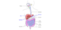
The digestive system takes in food, digests and absorbs nutrients, and eliminates the remaining waste material. The digestive system can be divided into the digestive tract (oral cavity, esophagus, stomach, small intestine, and large intestine) and associated digestive organs (salivary glands, pancreas, liver, and gallbladder).
Hard Palate
The hard palate is a thin horizontal bony plate located in the roof of the mouth that separates the oral and nasal cavities.
Tongue
The tongue is a moveable muscular process in the mouth that manipulates food for mastication and is used in the act of swallowing. The tongue's upper surface is covered by taste buds housed in numerous papillae.
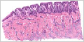

Esophagus
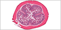
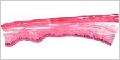
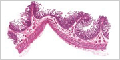
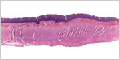
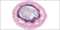
The esophagus is a muscular tube through which food passes from the mouth (pharynx) to the stomach. The muscular layer of the esophagus wall is composed of skeletal muscle in the upper part, smooth muscle in the lower part, and a mixture of the two in the middle.
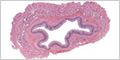

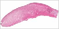
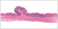
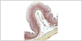
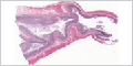
Gastroesophageal Junction
The gastroesophageal junction is the point where the distal esophagus joins the proximal stomach (cardia). It is the key defense against gastroesophageal reflux (GERD).
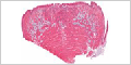
Stomach
The stomach digests food by acidification and the breakdown of proteins. It is divided into three histological regions (cardiac, body/fundus and pyloric) based on their anatomical location and appearance of their glands.
Cardiac stomach is the closest to the esophagus and contains mucus-secreting glands.
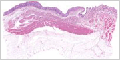
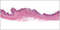
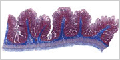
Fundic stomach is the largest part of the stomach (body/fundus) and contains fundic (gastric) glands.
Tissue sections from the fundic stomach stained with hematoxylin and eosin H&E.

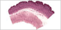
Pyloric stomach is closet to the small intestine and contains mucus-secreting glands.
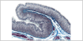
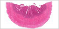
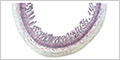
Vasculature of the Gastric Mucosa
The blood supply to the stomach wall can be seen after perfusion with a colored dye.
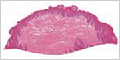
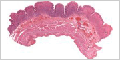
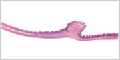
Gastroduodenal Junction
The gastroduodenal junction is the point where the distal stomach (pyloric) joins the proximal duodenum of the small intestine. The pyloric sphincter controls the passage of partially digested food ( i.e. , chyme) from the stomach into the duodenum
Small Intestine
The small intestine is involved in the continued digestion of food and the absorption of nutrients. It is divided into three segments: duodenum , jejunum , and ileum .


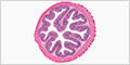
Vasculature of the Small Intestine
The blood supply to the small intestine can be seen after perfusion with a colored dye.
Large Intestine
The large intestine absorbs water and consolidates waste material into feces. It is divided into the cecum, appendix, colon, rectum and anal canal.


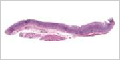
Rectum
The rectum is the final portion of the large intestine that acts as a temporary storage site for feces. Feces pass out of the rectum, through the anus, and out of the body.