Chapter 7 - Peripheral Blood
Blood is a fluid connective tissue composed of formed elements ( red blood cells , white blood cells , and platelets ) circulating in a fluid called plasma . It provides a mechanism by which gases, nutrients, wastes, and cells can be transported throughout the body.
Special stains are used to differentiate the types of blood cells. Wright's stain , a mixture of eosin Y (acidic) and methylene blue (basic) dyes, is superior in this regard to hematoxylin and eosin H&E.
Blood
Composition of human blood:
- 50 to 65% plasma
- 36 to 50% red blood cells (erythrocytes)
- < 1% white blood cells (leukocytes)
Composition of white blood cells :
-
Granulocytes
- contain azurophilic (primary) granules and specific (secondary) granules
- 60 to 70% neutrophils (polymorphonuclear leukocytes, PMNs)
- 2 to 4% eosinophils
- basophils
-
Agranulocytes
- contain only azurophilic (primary) granules
- 25 to 30% lymphocytes
- 5% monocytes
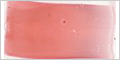
Blood Smear
The histology of blood cells is studied in smears prepared by spreading a drop of blood into a thin layer on a microscope slide.
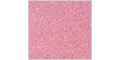
Blood Cells
The histology of blood cells is examined using a single, high-resolution 60x scan acquired from the zone of best morphology of this blood smear.
Identify the different types of blood cells by distinguishing characteristics, such as, size, shape, nuclear morphology, and the presence or absence of granules, and the appearance of granules.
Summary of blood cells identified on this slide.
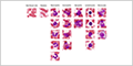
Review
This diagram allows the morphology and staining characteristics of different types of blood cells to be easily compared. The key features used to distinguish between them are highlighted.