Chapter 1 - The Cell
Histology is the study of the microscopic structure of cells, tissues, and organs, focusing on how cellular structure relates to biological function.
Objectives
The objective of this chapter is to develop fundamental skills needed to observe and analyze histological specimens using light microscopy. This chapter provides a diverse collection of cells, tissues, and organs to illustrate the variety of cellular forms found in humans.
It is not necessary to memorize specific cell and tissue names, but rather to develop observational skills for recognizing and interpreting cellular variations.
Characteristics to Observe and Notice
When examining histological specimens, focus on the following key cellular features:
- Size of the Cell : Compare cellular dimensions across different tissue types
- Shape of the Cell : Note morphological variations that reflect cellular function
- Nuclear/Cytoplasmic Ratio : Assess the proportion of nucleus to cytoplasm
- Chromatin Condensation : Distinguish between heterochromatin (condensed, transcriptionally inactive) and euchromatin (dispersed, transcriptionally active)
- Staining Properties : Identify basophilic or acidophilic staining patterns
- Secretion Granules : Observe specialized secretory structures
- Special Staining Properties : Note unique staining characteristics of specific organelles and cell types
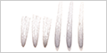
Light Microscope
Since most cells range from 10-30 µm in diameter - far below the resolution limit of the human eye (approximately 100 µm) — microscopes are essential tools for studying cellular structure and function. This microscopic investigation is fundamental to all biological sciences, as every process of life begins at the cellular level.
The light microscope remains the workhorse of histological investigation, providing magnifications upto 1000x with oil immersion objectives and achieving resolution limits of approximately 0.2 µm.
Histological Stains
Biological tissues present a unique challenge for microscopic examination - they are essentially transparent to visible light. The primary components of biological material (water, proteins, lipids, and carbohydrates) exhibit minimal absorption or scattering of light, making cellular structures virtually invisible under standard illumination.
Histological stains overcome this limitation by using specialized dyes and reagents that exploit differences in chemical properties - including electrical charge, hydrophobic associations, and enzymatic reactions — to bind selectively to distinct cellular and extracellular components. This selectivity in binding transforms invisible structures into clearly visible, color-coded architectural features.
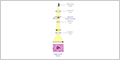
H&E Staining
The combination of hematoxylin and eosin (H&E) is the most widely used staining technique in histology and pathology. This pairing has dominated histology for over 150 years due to it's remarkable effectiveness in revealing cellular and tissue structure.
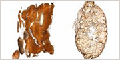
Hematoxylin is a basic dye that binds to acidic components, primarily nucleic acids (DNA and RNA),staining nuclei blue to purple. Eosin is an acidic dye that binds to basic components, particularly proteins in the cytoplasm, staining them pink to red. The complementary relationship between these dyes provides morphological detail that far exceeds the capabilities of either stain used alone.

The H&E staining of acinar cells can be correlated with it's ultrastructure revealed by transmission electron microscopy (TEM).
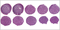
Cellular Diversity
All life on Earth, from the simplest bacteria to complex multicellular organisms like humans, is composed of cells . These cells display incredible diversity, varying dramatically in size, shape, and internal organization to fulfill specific roles within organisms.
The following examples illustrate a wide range of cellular morphologies and their corresponding staining patterns, demonstrating how form follows function in biological systems.
Specialized Histological Stains
While hematoxylin and eosin (H&E) staining provides excellent visualization of general cellular morphology, specialized stains can highlight specific cellular components and biochemical processes that remain invisible or poorly defined with routine stains.
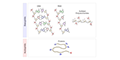
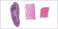
Toluidine Blue : This metachromatic dye selectively stains RNA-rich regions (such as ribosomes, rough endoplasmic reticulum, and nucleoli), in metabolically active cells. The dye exhibits metachromasia , appearing purple when bound to densely packed negatively charged groups like nucleic acids, while staining blue ( orthochromatic ) when bound to less concentrated negative charges.
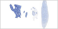
Golgi's Method : This classical silver impregnation technique, developed by Camillo Golgi, specifically visualize neurons and their processes with remarkable clarity. This method also stains the Golgi apparatus within other cell types.
Iron Hematoxylin : Certain formulations of this nuclear stain can be specifically adapted to visualize mitochondria, highlighting these crucial cellular organelles.

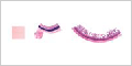
Feulgen Stain : This DNA-specific technique employs mild acid hydrolysis followed by reaction with Schiff's reagent to produce magenta-colored nuclei, while leaving RNA and proteins unstained.

Periodic Acid-Schiff (PAS) Reagent : This technique uses periodic acid to oxidize carbohydrate molecules followed by reaction with Schiff's reagent to produce magenta-colored deposits wherever glycogen and other carbohydrates are present.

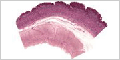
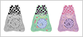
Purkinje Cells
Modern histological research rarely relies on a single staining technique. The complexity of cellular structure and function demands multiple complementary approaches to build a complete picture of biological processes.
Purkinje Cells in the cerebellum provide an ideal demonstration of how different staining techniques reveal complementary aspects of cellular structure and function. These large neurons, with their distinctive dendritic trees, showcase different cellular features depending on the staining method employed.
Mitosis
Animal Mitosis
Mitosis is the fundamental process during which a single parent cell divides to produce two genetically identical daughter cells. During this process, the chromosomes of the parent cell are duplicated and then systematically separated into the nuclei of the two daughter cells.
Mitosis consists of several distinct phases: prophase (chromosome condensation), metaphase (chromosome alignment), anaphase (chromosome separation), and telophase (nuclear reformation), followed by cytokinesis (cytoplasmic division).
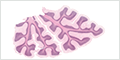
Plant Mitosis
Although onion root tips have been widely used as teaching examples of mitosis in histology courses due to their large, easily observable chromosomes and high mitotic activity, they present significant limitations for understanding mammalian cell biology.
Cytokinesis occurs through markedly different mechanisms in animal and plant cells, reflecting their distinct cellular architectures:
- Animal Cytokinesis : Actin and myosin form a contractile ring around the cell's middle that gradually constricts, pinching the plasma membrane inward, ultimately separating the two daughter cells through membrane fission.
- Plant Cytokinesis : Rather than pinching inwards, plant cells construct a new cell wall (called a cell plate) between the daughter nuclei. This structure is built from the center outward through the fusion of vesicles derived from the Golgi apparatus.