Chapter 8 - Hematopoiesis
Hematopoiesis is the development and formation of blood cells. Blood cells arise from hematopoietic stem cells rather than from the division of mature blood cells. This complex process involves the formation of many intermediate stages and cell types that become progressively more differentiated.
In adults, hematopoiesis occurs in bone marrow and generates hundreds of millions of new blood cells every day.
BONE MARROW
Bone marrow marrow is a specialized connective tissue composed of developing blood cells supported by reticular connective tissue. It exists in two distinct forms that differ in composition, function, and distribution:
-
Red bone marrow
:
- Red color in fresh tissue results from the large number of developing red blood cells and blood-filled sinusoids
- Site of production of blood cells throughout life (hematopoiesis)
- Predominantly found in flat bones (sternum, ribs, pelvis, vertebrae, skull) and the epiphyses of long bones in adults
- In infants and children, red marrow fills the medullary cavities of most bones
-
Yellow bone marrow
:
- Yellow color in fresh tissue results from the large numbers of adipose cells
- Adipose tissue replaces hematopoietic tissue in the medullary cavities of long bones during development
- Largely inactive in hematopoiesis under normal conditions but retains the capacity to revert to red marrow during periods of increased demand for blood cell production (such as severe blood loss and chronic anemia)

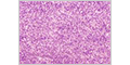
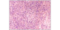
Bone Marrow Smear
The densely packed, highly cellular environment of bone marrow makes precise identification of individual cell types challenging in routine H&E-stained preparations. For detailed examination and classification of developing blood cells, bone marrow aspirate smears are the preferred method.
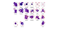
HEMATOPOIESIS
A high resolution scan from the zone of best morphology of a bone marrow smear was obtained using an oil immersion 60x lens (NA 1.4). The increased resolution allows the morphology and staining characteristics of the different cell types to be more easily examined.
Erythrocytes
Erythropoiesis is the development of red blood cells. As the precursor cells mature, they decrease in size, chromatin becomes more condensed, nuclei are eventually extruded, and cytoplasm changes in color from blue to pink. Granules are never seen in the lineage of red blood cells.
Granulocytes
Granulopoiesis is the development of granulocytes - neutrophils, eosinophils, and basophils. Key events in the maturation of granulocytes are the production of azurophilic granules, specific granules, and nuclear lobulation.
Agranulocytes
Lymphopoiesis and monopoiesis are the development of lymphocytes and monocytes, respectively. Unlike granulocytes, they do not contain specific granules or show nuclear lobulation.
Platelets
Thrombopoiesis is the development of platelets. The largest cells found in bone marrow are megakaryocytes which give rise to platelets.
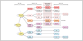
REVIEW
This diagram allows the morphology and staining characteristics of the different stages of blood cell development to be compared.
QUIZ
You should now be able to identify these different types of hematopoietic cells found in bone marrow.
PLASMA CELL
Bone marrow contains a survival niche that allows plasma cells to continuously secrete antibodies with a life span of months to years.
OSTEOCLAST
A rare example of an osteoclast can be seen in this bone marrow smear.

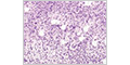
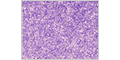
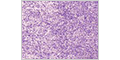
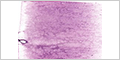
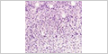
ADDITIONAL EXAMPLES
Viewing of these slides is not required. However, they demonstrate the variability seen between different preparations of bone marrow smears.