Chapter 14 - Gastrointestinal Tract
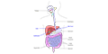
The digestive system takes in food, digests and absorbs nutrients, and eliminates the remaining waste material. The digestive system can be divided into the digestive tract (oral cavity, esophagus, stomach, small intestine, and large intestine) and associated digestive organs (salivary glands, pancreas, liver, and gallbladder).
STOMACH
The stomach digests food by acidification and the breakdown of proteins. It is divided into three histological regions (cardiac, body/fundus and pyloric) based on their anatomical location and appearance of their glands.

Fundic Stomach
Gastric glands are found in the fundus/body of the stomach and produce stomach acid (HCl) and secrete proteolytic enzymes.
Mucous neck cells are found in the upper parts (isthmus and neck) of the gastric glands. They are smaller than surface mucous cells and produce mucins. This mucus protects the epithelium from digesting itself.

Parietal cells produce hydrochloric acid (HCl) and release intrinsic factor, a glycoprotein essential for the absorption of vitamin B12 in the small intestine.

Chief cells are found at the base of gastric glands and secrete proteolytic enzymes (pepsinogen, chymosin, and gastric lipases)

Enteroendocrine cells are specialized secretory cells found in the simple columnar epithelium of the gastrointestinal tract. They release hormones from their basal surface that diffuse to nearby cells (paracrine) or into the bloodstream (endocrine).

Pyloric Stomach
Pyloric glands are located in the antrum of the pylorus.


GASTRODUODENAL JUNCTION
The gastroduodenal junction is the point where the distal stomach (pyloric) joins the proximal duodenum of the small intestine. The pyloric sphincter controls the passage of partially digested food ( i.e. , chyme) from the stomach into the duodenum

SMALL INTESTINE
The small intestine is where most of the digestion and absorption of nutrients occurs. It is divided into the duodenum , jejunum and ileum .
The surface area for absorption is increased 30-fold by broad, finger-like projections into the lumen of the small intestine.


The small intestine is lined with a simple columnar epithelium with goblet cells.

Intestinal Glands
The intestinal epithelium extends into the lamina propria to form intestinal glands (or intestinal crypts, crypts of Lieberkühn).


Blood Supply
The villi of the small intestine is richly supplied with blood to aid in the adsorption of nutrients.


Lacteals
Lacteals are blunt-ended lymphatic capillaries located at the center of villi in the small intestine.


Mucosa-Associated Lymphoid Tissue (MALT)
Lymphoid nodules become increasingly numerous in the ileum and form bulges called Peyer’s patches . The epithelium that covers Peyer's patches contain specialized epithelial cells (M cells) that transport antigens to immune cells to initiate an immune response.


ILEOCECAL JUNCTION
The ileocecal junction is the boundary between the small intestine and the large intestine.

LARGE INTESTINE
The large intestine absorbs water and consolidates the fecal mass. It is divided into the cecum, appendix, colon, rectum and anal canal.


