Chapter 2 - Epithelium
Epithelium forms continuous sheets of cells that line internal surfaces and cover the external surface of the body. It acts as a selective barrier that protects tissues. A basement membrane separates the epithelium from underlying connective tissue.
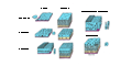
Epithelium is classified based on three criteria:
- Number of cell layers ( single or compound )
- Shape of surface cells ( squamous , cuboidal or columnar )
- Specializations ( cilia , keratin or goblet cells )
Epithelial cells are polarized:
-
Apical domain
- surface that faces the lumen or the external environment
- Microvilli (brush border), cilia, stereocilia
-
Lateral domain
- surfaces that face the sides of adjacent cells
- Tight junctions (zonula occludens), adherens junction (zonula adherens), desmosomes (macula adherens), gap junctions
-
Basal domain
- surface that attaches to the basement membrane
- Basement membrane, hemidesmosomes
Epithelium does not contain blood vessels and receives nourishment via diffusion from the underlying connective tissue.
Glands are formed by the down growth of an epithelium into the underlying connective tissue.
It is not necessary to learn the names of specific tissues for this chapter, but rather learn to recognize variations in epithelia.
Simple Squamous Epithelium
Simple squamous epithelium consists of a single layer of flattened cells in contact with the basement membrane. The thinness of these cells facilitates the selective transfer of materials ( e.g. , gases, fluids or nutrients) across the epithelium.


Simple Cuboidal Epithelium
Simple cuboidal epithelium consists of a single layer of cuboidal cells. This epithelium is often associated with absorption, secretion or excretion of waste matter.

Simple Columnar Epithelium
Simple columnar epithelium consists of a single layer of cells that are taller than they are wide. This epithelium is often associated with absorption or secretion.
Small Intestine
Simple columnar epithelium with goblet cells of the small intestine.
Gallbladder
Simple columnar epithelium of the gallbladder.


Pseudostratified Columnar Epithelium
Pseudostratified columnar epithelium appears to be stratified because the nuclei of the epithelial cells are at different levels, but every cell is in contact the basement membrane. The epithelial cells vary in height.


Stratified Squamous Epithelium
Stratified squamous epithelium has multiple layers of cells becoming flattened as they move from the basal layer to the apical layers. It provides protection from abrasion and is keratinized on the external surface of the body.

Microvilli
Microvilli are cellular extensions on epithelial cells that increase the surface area of the apical plasma membrane.

Cilia
Cilia are slender, hair-like appendages that extend from the surface of cells. There are two types of cilia on human cells:
- Non-motile cilia (primary cilia) - found on most cells and usually serve as sensory organs.
- Motile cilia - found in large numbers on some cells and beat in coordinated waves that move material across the surface of an epithelium.


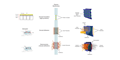
Intercellular Junctions
Intercellular junctions are specialized structures that connect adjacent cells, enabling communication, adhesion, and coordination within tissues.
- Tight Junctions (zonula occludens) - seal the space between adjacent cells forming a barrier to diffusion
- Adherens Junctions (zonula adherens) - ribbon-like structures that provide mechanical attachment between adjacent cells
- Desmosomes (macula adherens) - spot-like adhesions that provide resistance to mechanical stress
- Gap junctions - aqueous channels between the cytoplasm of adjacent cells that allow the passage of small molecules
The small intestine is lined by tightly packed, simple columnar epithelial cells that form a selective barrier, facilitating nutrient absorption while protecting the underlying tissues.

The junctional complex of the small intestine epithelium, also known as the terminal bar under light microscopy, is a specialized region at the apical end of epithelial cells that plays a critical role in maintaining intestinal barrier function and cell polarity.


Skin cells (keratinocytes) contain numerous adherens junctions (zonula adherens) which tightly join cells in the epidermis. They provide mechanical resistance to physical stress helping maintain structural integrity under stretching, pressure, and friction.


Gap Junctions are specialized intercellular connections formed by protein channels known as connexons. They enable the rapid and regulated exchange of signaling molecules (such as Ca 2+ and cAMP) between neighboring cells, allowing groups of cells to coordinate their response.
