Chapter 9 - Cardiovascular System
The cardiovascular system transports blood to and from the heart to all tissues of the body. Its main function is to transport oxygen and carbon dioxide, nutrients, and metabolic waste products. It is also involved in temperature regulation, hormone distribution, and immune function.
The cardiovascular system is composed of the following structures:
- Heart - pumps blood through the system
- Arteries - vessels that deliver blood to tissues
- Capillaries - networks of small vessels that perfuse tissues
- Veins - vessels that return blood to the heart
Blood vessels are found in all tissues of the body with very few exceptions, such as epithelia and cartilage.
Blood cells are examined in a separate chapter.
HEART
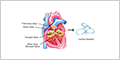
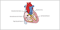
The heart is a four-chambered muscular organ consisting of two upper chambers called atria (right and left) that receive blood, and two lower chambers called ventricles (right and left) that pump blood out of the heart. The right side of the heart receives deoxygenated blood from the body and pumps it to the lungs, while the left side receives oxygenated blood from the lungs and pumps it to the rest of the body.
Cardiac muscle , also known as myocardium, is a type of striated, involuntary muscle tissue that is exclusively found within the heart wall. It combines the organized contractile apparatus of skeletal muscle with the autonomous function of smooth muscle.

The cardiac atria are the two upper chambers of the heart, serving as low-pressure receiving chambers that collect blood from both the systemic and pulmonary circulations.

The cardiac ventricles serve as the high-pressure pumps that propel blood through the pulmonary and systemic circulations.


Heart Valves and Cardiac Skeleton
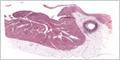
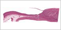
Heart valves are thin folds of the endocardium with a core of dense connective tissue. To maintain their shape, heart valves are attached around the fibrous rings of the cardiac skeleton .
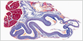
Purkinje Fibers
The cardiac conduction system transmits signals that coordinate muscle contractions, pumping blood throughout the circulatory system. Electrical signals generated by the sinoatrial node, the heart's pacemaker, travel through the right atrium to reach the Purkinje fibers in the ventricular walls.


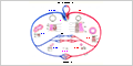
BLOOD CIRCULATION
Blood circulation is divided into two circuits:
- Pulmonary circuit - carries oxygen-poor blood from the heart to the lungs and returns as oxygen-rich blood (see Respiratory System )
- Systemic circuit - carries oxygen-rich blood from the heart throughout the body and returns as oxygen-poor blood
Blood vessels have walls composed of three layers (or tunics):
- Tunica intima - endothelium and loose connective tissue
-
Tunica media
- concentric layers of varying amounts of elastic fibers, smooth muscle cells, and collagen fibers
- Typically the thickest layer in arteries
-
Tunic adventitia
- outer layer of connective tissue
- Typically the thickest layer in veins
These layers vary in thickness depending on the type of vessel (arteries, arterioles, venules, and veins).
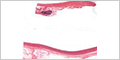
Elastic Arteries and Large Veins
Elastic arteries conduct blood from the heart to different areas of the body. These vessels include the aorta, pulmonary artery, and their largest branches.
The tunica media contains many concentric fenestrated sheets of elastin ( i.e. , elastic laminae ) interspersed with smooth muscle cells.
This elastic tissue allows these vessels to distend when the blood pressure rises (systole), and recoil when the blood pressure fails (diastole). This pumping action helps maintain blood pressure through the cardiac cycle.


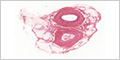
Muscular Arteries and Medium Veins
Muscular arteries distribute blood to specific organs in response to their functional needs. Most of the named arteries in the body are muscular arteries.
The tunica media is composed of concentric layers of smooth muscle cells. The contraction and relaxation of these muscle cells regulate blood flow by changing the size of the lumen.
A prominent internal elastic lamina separates the tunica intima from the media. In larger arteries, an external elastic lamina also separates the tunica media from the adventitia.

Arterioles and Venules
Arterioles regulate the flow of blood into capillary beds. They provide the majority of the resistance to blood flow in the body.
The tunic media is reduced to one or two concentric layers of smooth muscle cells.
The contraction of the smooth muscle cells constricts the lumen of the arteriole, reducing the flow of blood, and increasing vascular resistance.
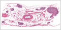
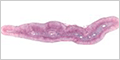
Capillaries
Capillaries are the smallest blood vessels (often less than 10 µm in diameter). The thin wall of capillaries is composed of endothelial cells supported by a basement membrane.
Three types of capillaries can be distinguished:
- Continuous capillaries - continuous endothelium and basement membrane
-
Fenestrated capillaries
- endothelial cells contain fenestrations, or pores, 80 to 100 nm in diameter (typically with thin diaphragms) only visible with
electron microscopy
- Rapid bidirectional movement of hormones, nutrients, and small molecules while preventing cellular passage
- Found in organs involved in filtration, secretion, or absorption of molecules - including endocrine glands, the kidneys, and the intestinal villi
-
Sinusoidal capillaries
- discontinuous endothelium and incomplete basement membrane
- Facilitate the movement of cells between the circulation and tissues
- Found in the liver, spleen, and bone marrow
Continuous capillaries are responsible for the exchange of gases, nutrients, and waste products between blood and tissues. Gases pass through endothelial cells by diffusion, small molecules pass between endothelial cells, while large molecules are transported across the endothelium by pinocytotic vesicles.
Fenestrated capillaries are typically found in organs involved in filtration, secretion, or absorption of molecules - including endocrine glands, the kidneys, and the intestinal villi. The pores increase permeability compared to continuous capillaries.
Capillary beds are an interconnected network of capillaries that perfuse organs and tissues.
Venous Valve
Medium and large veins have valves that prevent the retrograde flow of blood.
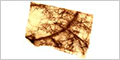
ATHEROSCLEROSIS
Atherosclerosis can result from injury to the tunica intima by hypertension, bacterial or viral infections, or chemicals in the blood. This damage results in the thickening of the tunica intima.