Chapter 4 - Muscle Tissue
Muscle tissue is composed of cells specialized for contraction. Muscle is classified into three types according to their structure and function:
- Skeletal muscle cells - striated, voluntary control
- Cardiac muscle cells - striated, involuntary control
- Smooth muscle cells - nonstriated, involuntary control
Skeletal and cardiac muscle cells are called striated because they show an alternating series of bands. The repeating arrangement of their basic contractile unit, the sarcomere , produces these striations.
In all types of muscle, contraction is caused by the movement of myosin filaments along actin filaments .
The terms muscle cell and muscle fiber are synonymous.
Skeletal Muscle
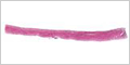
Skeletal muscle fibers are long cylindrical, multinucleated, striated, and under voluntary control.
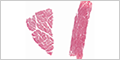


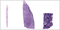
Individual skeletal muscle cells can be seen by teasing apart a muscle.

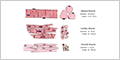
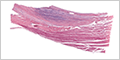
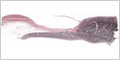
Muscle Bone Junctions
Muscles connect to the skeletal framework to enable motion and provide stability. These connections transmit the forces generated by muscle fibers (cells) through two distinct mechanisms:
- Tendons - muscles taper at their ends into strong, fibrous cords or sheet-like structures that connect muscle to bone
- Muscle Insertions - collagen fibers of the endomysium (connective tissue surrounding individual muscle cells) extend and interweave with the collagen fibers of the periosteum covering bone.


Cardiac Muscle
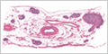
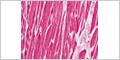
Cardiac muscle are short branching fibers, have a single, centrally located nucleus, show the same striations as skeletal muscle, and are under involuntary control.
Purkinje fibers are modified cardiac muscle cells that convey electrical impulses that coordinate contraction of cardiac muscle.

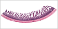
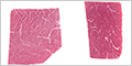
Smooth Muscle
Smooth muscle cells are spindle-shaped (fusiform), have a single, centrally located nucleus, and are under involuntary control. The uniform, nonstriated appearance gives rise to the name smooth muscle.