Faculty/Retired
University of Minnesota
Department of Genetics, Cell Biology and Development
6-160 Jackson Hall
321 Church St SE
Minneapolis, MN 55455
Robert L. Sorenson, Ph.D.
Professor Emeritus
University of Minnesota
Department of Genetics, Cell Biology and Development
6-160 Jackson Hall
321 Church St SE
Minneapolis, MN 55455
Download the User Guide v1.1 (PDF) to learn about new platform features.
Each slide is shown with additional information to its right. The image can be changed using any combination of the following commands.
Sidebar
Links: Click to navigate to a specific region
Images: Click to show this view
Toolbar: Use controls to adjust magnification and pan the image
Mouse
Zoom In: Click left button
Zoom Out: Double-click left button
Pan/Move: Click and drag the image
Keyboard
Zoom In: ‘A’ key
Zoom Out: ‘Z’ key
Pan/Move: Arrow keys (Up, Down, Left, Right)
Reset View: ESC key (fit-to-screen view)
Touch
Tap: Zoom in on a specific area
Double-tap: Zoom out from the current view
Drag: Pan the image
SHARE
A link to a virtual slide can be saved for later viewing in different ways.
Clipboard
The address of this view has been copied to your clipboard. This link can be pasted in any other program.
Bookmark
A bookmark link can be created using the bookmark function (Ctrl-D for Windows or Cmd-D for Mac) of your browser. Choose a name for the bookmark and select the folder in which you want it saved.
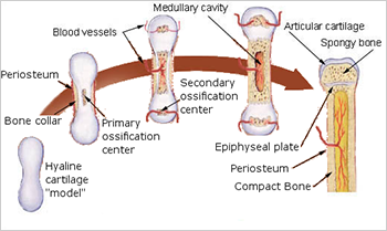
MHS 287 Bone
Endochondral Bone Development
Endochondral ossification is a process where bone replaces cartilage. The various stages can be seen in these front and rear legs of a fetal rat.
The upper shows the early stages of the formation of primary centers of ossification.
form in the approximate shape of the adult bone.
Steps in the formation of :
Periosteum forms a bony collar in the diaphyseal region of the cartilage
Chondrocytes show early hypertrophy
Chondrocytes calcify their matrix and die
Osteoblasts differentiate from the periosteum and begin to lay down the
Endochondral Bone Development
The lower shows the later stages of bone development. The shows the formation of the epiphyseal plates.
The can be seen in the diaphysis.
The epiphyseal plates (,) continuously provide for the lengthening of the bone.
(Reserve) - quiescent chondrocytes
- rapidly dividing chondrocytes that form longitudinal columns of stacked cells
- chondrocytes cease dividing, grow in size, and secrete cartilage matrix
- calcification of the cartilage matrix inhibits diffusion of nutrients and the chondrocytes undergo apoptosis
- osteoprogenitor cells migrate into the cavities with new blood vessels, differentiate into osteoblasts, and deposit (eosinophilic) on the scaffold of calcified cartilage (basophilic)