Martin J.C. Dane 1,3 Bernard M. van den Berg 1 M. Cristina Avramut 1 Frank G.A. Faas 1 Johan van der Viag 3 Angelique L.M.M.M. Rops 3 Raimond B.G. Ravelli 1 Bram J. Koster 1 Anton Jan van Zonnevekd 1 Hans Vink 2 Ton J. Rabelink 1
1Leiden University Medical Center
Leiden, Netherlands
2Maastricht University Medical Center
Maastricht, Netherlands
3Nijmegen Medical Center
Radboud University
Nijmegen, Netherlands
Faculty/Retired
University of Minnesota
Department of Genetics, Cell Biology and Development
6-160 Jackson Hall
321 Church St SE
Minneapolis, MN 55455
Robert L. Sorenson, Ph.D.
Professor Emeritus
University of Minnesota
Department of Genetics, Cell Biology and Development
6-160 Jackson Hall
321 Church St SE
Minneapolis, MN 55455
Download the User Guide v1.1 (PDF) to learn about new platform features.
Each slide is shown with additional information to its right. The image can be changed using any combination of the following commands.
Sidebar
Links: Click to navigate to a specific region
Images: Click to show this view
Toolbar: Use controls to adjust magnification and pan the image
Mouse
Zoom In: Click left button
Zoom Out: Double-click left button
Pan/Move: Click and drag the image
Keyboard
Zoom In: ‘A’ key
Zoom Out: ‘Z’ key
Pan/Move: Arrow keys (Up, Down, Left, Right)
Reset View: ESC key (fit-to-screen view)
Touch
Tap: Zoom in on a specific area
Double-tap: Zoom out from the current view
Drag: Pan the image
SHARE
A link to a micrograph can be saved for later viewing in different ways.
Clipboard
The address of this view has been copied to your clipboard. This link can be pasted in any other program.
Bookmark
A bookmark link can be created using the bookmark function (Ctrl-D for Windows or Cmd-D for Mac) of your browser. Choose a name for the bookmark and select the folder in which you want it saved.
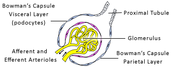
EM 364 Renal Corpuscle
Renal Corpuscle
Transmission electron micrograph (TEM) of a renal corpuscle in a kidney.
Electron microscopy enables high-resolution examination of tissue, but it can be challenging to observe large structures. This remarkable micrograph of a renal corpuscle is an exceptionally large montage of 81 x 76 images (6,156 images) covering an area of 144 x 134 µm.
Glomerulus
(red) - lumen (pink)
(blue and green)
(orange)/Matrix (tan)
(brown)
(tan, upper right) - part of the initial segment
Cell Structures
Nuclei (blue) / Nuclear Envelope (purple)
Golgi Apparatus (yellow)
Mitochondria (red)
Endoplasmic Reticulum (cyan)
Basal Lamina (purple)
Courtesy of Bernard van den Berg and Antonius Johannes Rabelink, Department of Internal Medicine, Leiden University Medical Center, Leiden, Netherlands.
Click the Information button in the toolbar for the complete credits.
Glomerulus
The is a tuft of capillaries that extends into the sac formed by the terminal end of the renal tubule. This cup-shaped structure is called Bowman's capsule.
A single afferent arteriole enters the glomerulus, forms about 20 to 40 capillary loops (red cells with pink lumen) and then emerges as an afferent arteriole. The point where the blood vessels enter and exit the glomerulus is known as the vascular pole of the renal corpuscle.
Bowman's capsule is composed of two main components:
Parietal Layer (,) – outer layer composed of a simple squamous epithelium (brown) continuous with renal tubule.
Visceral Layer (,) – inner layer composed of a thin, specialized layer of epithelial cells called podocytes (blue and green).
The exit point of Bowman's capsule into the renal tubule is known as the urinary pole of the renal corpuscle.
Glomerular Filtration
The glomerulus is a tuft of capillaries where water, ions, and small molecules are filtered out of the blood into the space made by Bowman's capsule.
The permeability barrier for filtering plasma is made up of three closely apposed parts:
Fenestrated Capillaries (, ,; red cells with pink lumen) - lined by endothelial cells with numerous pores (or fenestrations) with an average diameter of 70 nm.
Filters plasma from blood
Fenestrations (, ,) lack diaphragms
Some red blood cells (dark red) and white blood cells (gray) are seen in the lumens of some capillaries
Basement Membrane (, ,; purple) - sandwiched between the capillaries and podocytes
Barrier to plasma proteins (such as albumin)
Formed by the endothelial cells and adjacent podocytes
Much thicker (300 to 400 nm) than in most tissues
Podocytes (,; blue and green) - highly specialized epithelial cells with long processes that wrap around the capillary loops (i.e., the visceral layer of Bowman's capsule).
Each podocyte has several primary branches, which give rise to many secondary processes (pedicles or foot processes).
Pedicles (,) of adjacent podocytes interdigitate and form filtration slits about 20 to 25 nm wide
Thin, non-membranous diaphragms span each filtration slit
The podocyte slit diaphragm is the main structure that controls the permeability of the glomerulus. Small molecules, such as glucose, can easily pass through, while larger molecules like albumin (69 kDa) can barely cross the barrier. Negatively charged, large molecules pass through less easily compared to positively charged ones of the same size.
Mesangium
The mesangium is a connective tissue framework that provides structural support to the tuft.
Mesangial Cells (,; orange) - pericyte-like cells located between the capillary loops
Phagocytic role in maintaining the basement membrane
Can contract to regulate blood flow in glomerular capillaries
Mesangial Matrix (tan) - extracellular matrix secreted by mesangial cells
Immune complexes may be phagocytosed by mesangial cells and, together with increased mesangial matrix production, may contribute to glomerular dysfunction.
Extraglomerular mesangial cells form an outward extension at the vascular pole and are a component of the juxtaglomerular apparatus (not visible).
Proximal Convoluted Tubule
Proximal Convoluted Tubules arise from the urinary poles of the Bowman's capsule. They are highly convoluted in the cortex and are sectioned randomly in circular, elliptical, or U-shaped profiles.
A small segment of the (tan, upper right) is seen exiting the renal corpuscle at the urinary pole.
Proximal convoluted tubules have a with a brush border of tall , reflecting its role in reabsorption and transport.
60 to 70% of the glomerular filtrate
Most of the water, sodium, glucose, amino acids, potassium, and citrate
Regulates the pH of the filtrate by secreting H+ into the lumen and reabsorbing bicarbonate
- arise from canaliculi in the apical cytoplasm that reabsorb and digest proteins
Proteins digested to amino acids
Basal Infoldings (,) - extensive deep invaginations into the cell's cytoplasm, which greatly increases the surface area compared to a non-folded membrane
Mitochondria packed between infoldings create a pattern of basal striations
Allows close proximity of plasma cell membrane and mitochondria
Absorbed molecules and amino acids are transported out of the cell into the peritubular capillaries